Ultrasound vs. X-ray: When Do We Use Each?
- Ella Riley CertNCS (VCC), RVCCA

- Oct 31
- 2 min read
Understanding Diagnostic Imaging in Equine Practice
Two of the most commonly used imaging tools are X-rays and ultrasound. While both are essential for diagnosing injury and illness in horses, they’re used to assess very different types of tissues — and knowing when to use each allows us to make accurate, informed decisions about your horse’s care.
Here’s a breakdown of what each tool does, when we use them, and why it matters.
What Are Radiographs (X-rays)?
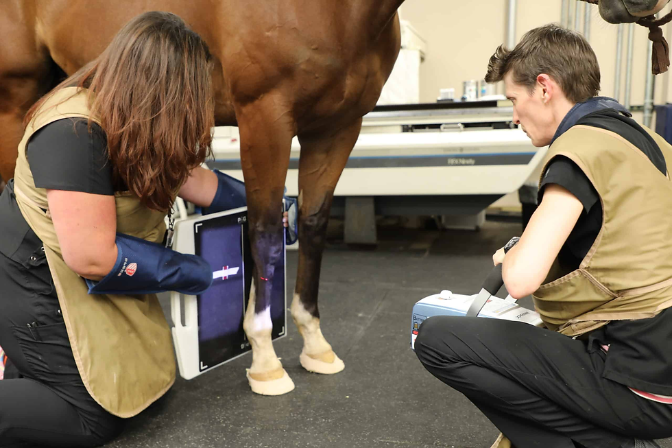
X-rays use a small amount of radiation to create images of dense tissues, such as bone.
What X-rays are best for:
Bone injuries (fractures, chips, bone bruising)
Joint conditions (arthritis, OCD, bone cysts)
Hoof balance and conformation
Dental disease
Pre-purchase assessments
Because X-rays show changes in bone density and structure, they’re not suitable for imaging tendons, ligaments, or soft tissue swelling — but they’re ideal for identifying bone abnormalities, even subtle ones.
What Is Ultrasound?
Ultrasound uses high-frequency sound waves to produce real-time images of soft tissue structures. It’s non-invasive, radiation-free, and can be performed repeatedly to monitor healing over time.
What ultrasound is best for:
Tendon and ligament injuries (e.g. SDFT, suspensory ligament)
Joint effusion and inflammation
Reproductive exams
Chest and abdominal imaging
Guided injections or aspiration of fluid
Monitoring healing after injury
Ultrasound excels at showing soft tissue texture, fibre alignment, and fluid-filled structures — but can’t penetrate bone, so it won’t show fractures or deeper bone lesions.
So When Do We Use Each?
Here’s a simple comparison to help:
Condition | X-ray | Ultrasound |
Fracture | ✅ Yes | ❌ No |
Arthritis | ✅ Yes | ❌ No |
Tendon injury | ❌ No | ✅ Yes |
Joint swelling | 🔄 Sometimes | ✅ Yes |
Hoof pain | ✅ Yes | 🔄 Sometimes (for navicular bursa) |
Lumps & bumps | ❌ No | ✅ Yes |
Breeding work | ❌ No | ✅ Yes |
Abdominal concerns | ❌ No | ✅ Yes |
In many cases, we use both tools together. For example, if a horse presents with a swollen joint and lameness, X-rays might rule out bone involvement, while ultrasound helps us assess the soft tissue structures around the joint.
How This Helps Your Horse
By using the right imaging tool for the job, we can:
Make faster, more accurate diagnoses
Avoid unnecessary treatments
Monitor progress over time
Tailor treatment plans for better outcomes
Imaging also helps us identify subtle or hidden injuries before they become more serious.
We're Here to Help
If your horse is lame, sore, or just not performing as they should, diagnostic imaging may be the next step. Whether it's X-rays, ultrasound, or both, our vets will guide you through the process with care and clarity.


Comments